Home » Without Label » Muscles Of The Chest Abdomen : Dont'a Hightower's Pec Tear | Family Physical Therapy Services / Fabian identifying the muscles and landmarks of the abdomen and chest.
Muscles Of The Chest Abdomen : Dont'a Hightower's Pec Tear | Family Physical Therapy Services / Fabian identifying the muscles and landmarks of the abdomen and chest.
Muscles Of The Chest Abdomen : Dont'a Hightower's Pec Tear | Family Physical Therapy Services / Fabian identifying the muscles and landmarks of the abdomen and chest.. There is a printable worksheet available for download here so you can take the quiz with pen and paper. Lying exposed between the protective bones of the superiorly located ribs and the inferiorly located pelvic girdle, the muscles of this region play a critical role in protecting the. One of the main smooth muscles inside the chest is the diaphragm. Muscles of the chest and abdomen— presentation transcript 24 muscles that move the arm (3 of 3) pectoralis major: Muscles abdomen & chest, back muscles label, muscles of the back.
Chest muscles are responsible for adduction, internal rotation, and forwards flexion of the humerus. Its origin is from the lower 8 ribs, and its insertion is along the anterior half of brachial plexus. Fabian identifying the muscles and landmarks of the abdomen and chest. Back muscles chart 12 photos of the back muscles chart back muscles chart, back muscles diagram and ligaments, back muscles diagram lats, back muscles diagram massage, upper back muscles chart, human muscles, back muscles chart, back muscles diagram and ligaments, back muscles diagram lats, back muscles diagram massage. If you've pulled a muscle—particularly in your chest, abdomen, or upper/middle back area—you may experience chest tightness and pain when you engage in activities.
Quotes about Abdomen (46 quotes) from www.quotemaster.org This may also radiate to the shoulder, arm, or even cause abdominal discomfort. The rectus abdominis muscle, also known as the abs and lower abdominals, is a paired muscle running vertically on each side of the anterior wall of the human abdomen. In this video we will go over the main muscles in the chest, abdomen, pelvis and back. Identify the movement and function of the intrinsic skeletal muscles of the back and neck, and the skeletal muscles of the abdominal wall and thorax. The skeletal muscles of the abdomen form part of the abdominal wall, which holds and protects the gastrointestinal system. Abdominal muscle, any of the muscles of the anterolateral walls of the abdominal cavity, composed of three flat muscular sheets, from. The muscles of the abdomen, lower back, and pelvis are separated from those of the chest by the muscular wall of the diaphragm, the critical breathing muscle. Muscles of the chest and abdomen learn by taking a quiz;
Starting with the rhomboid muscle divided into major and minor and con.
Learn vocabulary, terms, and more with flashcards, games, and other study tools. If you've pulled a muscle—particularly in your chest, abdomen, or upper/middle back area—you may experience chest tightness and pain when you engage in activities. One of the main smooth muscles inside the chest is the diaphragm. Fabian identifying the muscles and landmarks of the abdomen and chest. The rectus abdominis muscle, also known as the abs and lower abdominals, is a paired muscle running vertically on each side of the anterior wall of the human abdomen. It plays a crucial role in the respiratory system by helping a person breathe. Originates from the upper portion of the sternum. The largest muscle and the one that brings the most concern is the heart. The pelvic floor digital book. Moving down the trunk of the cat from the chest to the abdomen, i was able to identify the latissimus dorsi, internal oblique, transverse abdominus, rectus abdominus, linea alba, and external oblique. The diaphragm forms the upper surface of the abdomen. The abdomen (commonly called the belly) is the body space between the thorax (chest) and pelvis. Back muscles chart 12 photos of the back muscles chart back muscles chart, back muscles diagram and ligaments, back muscles diagram lats, back muscles diagram massage, upper back muscles chart, human muscles, back muscles chart, back muscles diagram and ligaments, back muscles diagram lats, back muscles diagram massage.
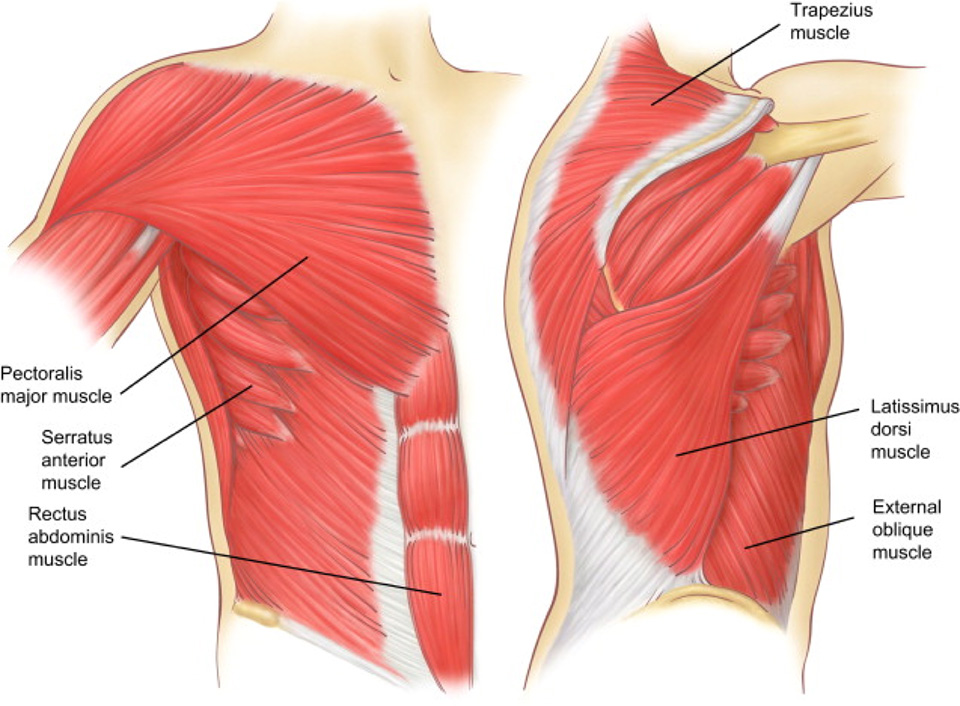
The muscles of the vertebral column, thorax, and abdominal wall extend, flex, and stabilize different parts of the body's trunk. The rectus abdominis is positioned between the ribs and the pubic bone at the front of the pelvis, and is actually made up of 8 distinct muscle bellies. Abdominal head of pectoralis major The pectoralis major, the pectoralis minor, and the serratus anterior. Identify the movement and function of the intrinsic skeletal muscles of the back and neck, and the skeletal muscles of the abdominal wall and thorax.
Axial Muscles of the Abdominal Wall and Thorax | Anatomy ... from s3-us-west-2.amazonaws.com One of the most common symptoms of pulling a chest muscle is pain around the affected muscle. Each one spans half of the upper chest, and has attachment points on the sternum (breastbone), ribs, clavicle (collarbone), and humerus. William blahd on webmd says that pulled muscle, strains, and tears can damage the muscle fibers and tendons. The chest wall is full of muscles, just like the arms and legs. In some cases, the strain may be severe enough to cause pain when breathing. The skeletal muscles of the abdomen form part of the abdominal wall, which holds and protects the gastrointestinal system. Muscles of the chest enable us to lift, extend, and rotate our arms, along with playing a part in the process of respiration. Learn vocabulary, terms, and more with flashcards, games, and other study tools.
Muscles abdomen & chest, back muscles label, muscles of the back.
.e193 pain in the thorax or abdomen can be the result of a local. Chest pain can be the result of pulled, strained, or sprained muscles in the chest or between the ribs. Fabian identifying the muscles and landmarks of the abdomen and chest. Bruising of the chest wall. If you've pulled a muscle—particularly in your chest, abdomen, or upper/middle back area—you may experience chest tightness and pain when you engage in activities. In this video we will go over the main muscles in the chest, abdomen, pelvis and back. Originates from the upper portion of the sternum. The diaphragm forms the upper surface of the abdomen. At the level of the pelvic bones, the abdomen. The skeletal muscles of the abdomen form part of the abdominal wall, which holds and protects the gastrointestinal system. Inserts along almost the entire length of the humerus and on the fascia covering the proximal end of the forearm. Of the two chest muscles, the pectoralis major (a.k.a. The muscles of the abdomen, lower back, and pelvis are separated from those of the chest by the muscular wall of the diaphragm, the critical breathing muscle.
The external oblique muscle is a broad muscle that runs along the anterolateral abdomen and chest wall. The pectoralis major, the pectoralis minor, and the serratus anterior. Muscles of the chest enable us to lift, extend, and rotate our arms, along with playing a part in the process of respiration. The pelvic floor digital book. At the level of the pelvic bones, the abdomen.
Costochondritis & Chest Wall Pain | Rib Injury Clinic from www.ribinjuryclinic.com The diaphragm forms the upper surface of the abdomen. The external oblique muscle is a broad muscle that runs along the anterolateral abdomen and chest wall. Muscles of the chest and abdomen learn by taking a quiz; The diaphragm is a muscle that acts as a partition between the upper abdomen and the chest. Muscles abdomen & chest, back muscles label, muscles of the back. Back muscles chart 12 photos of the back muscles chart back muscles chart, back muscles diagram and ligaments, back muscles diagram lats, back muscles diagram massage, upper back muscles chart, human muscles, back muscles chart, back muscles diagram and ligaments, back muscles diagram lats, back muscles diagram massage. Disorders of the inert structures. Inserts along almost the entire length of the humerus and on the fascia covering the proximal end of the forearm.
Any injury to your chest can cause chest pain.
Any injury to your chest can cause chest pain. Spasms can also occur when a muscle is overused, injured, tired, or strained. Each one spans half of the upper chest, and has attachment points on the sternum (breastbone), ribs, clavicle (collarbone), and humerus. The largest muscle and the one that brings the most concern is the heart. Identify the movement and function of the intrinsic skeletal muscles of the back and neck, and the skeletal muscles of the abdominal wall and thorax. Chest pain can be the result of pulled, strained, or sprained muscles in the chest or between the ribs. The external oblique muscle is a broad muscle that runs along the anterolateral abdomen and chest wall. Bruising of the chest wall. Draws scapula laterally and forward around chest wall; Muscle diagram free weights muscle anatomy chest muscles nursing tips medical science upper body back pain scorpio. The pelvic floor digital book. Originates from the upper portion of the sternum. Back muscles chart 12 photos of the back muscles chart back muscles chart, back muscles diagram and ligaments, back muscles diagram lats, back muscles diagram massage, upper back muscles chart, human muscles, back muscles chart, back muscles diagram and ligaments, back muscles diagram lats, back muscles diagram massage.